Nuclear medicine is a medical specialty which uses safe, painless and cost-effective techniques both to image the body and treat disease.
Nuclear medicine imaging is unique in that it documents organ function, structure and quantification, in contrast to diagnostic radiology which is based upon anatomy. It is a way to gather medical information that may otherwise be unavailable, require surgery, or necessitate more expensive diagnostic tests.
As an integral part of patient's care, nuclear medicine is used in the diagnosis, treatment and prevention of serious disease. Nuclear medicine imaging procedures often identify abnormalities very early in the progression of a disease –long before some medical problems are apparent with other diagnostic tests. This early detection allows a disease to be treated early when there may be a more successful prognosis.
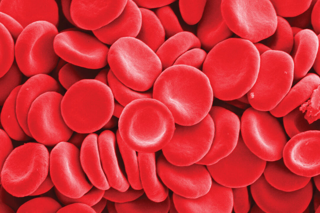
Nuclear medicine uses very small amounts of radioactive materials to diagnose and treat disease. These radiopharmaceuticals are substances that are attracted to specific organs, bones, or tissues. When introduced into the body, they produce gamma emissions. A provide information about the area of the body being imaged.
Although Nuclear Medicine is commonly used for diagnostic purposes, it also provides valuable therapeutic applications such as treatment of hyperthyroidism, thyroid cancer, types of bone cancer. Today, there are nearly 100 different nuclear medicine imaging procedures which provide information about virtually every major organ system within the body. Nuclear medicine now is an integral part of patient's care, and an important diagnostic and therapeutic specialty in medical science. Clinical applications Nuclear medicine imaging uniquely provides information about both the function and structure of virtually every major organ system within the body. Some of common applications are as follows :
Nuclear Endocrinology:
A. Thyroid isotope scan:
B. Parathyroid isotope scan:
C. Drenal gland isotope scan:
Kidney Isotope Scan:
A. Dynamci renal scan and diuretic renagram (DTPA, -MAG-3) B. Static renal scan (DMSA):
Nuclear Cardiology:
A. Myocardial Stress- Rest perfusion scan (Sestamibi, Thallium201):
B. Gated Blood pool study (MUGA):
Gastrointestinal isotope scan:
A. Liver – spleen scan:
B. Hepato-biliary (HIDA) scan:
C. Liver blood pool scan ( labeled RBCs):
D. G.I Bleeding scan ( labeled RBCs)
E. Gastric emptying study
F. Ectopic gastric mucosa scan:
G. Salivary gland scan:
Lymphoscintgraphy:
Epididymo-orchitis:
Tumor imaging:
A. Gallium -67 scan:
B. Thallium -201, Sestumibi (MIBI):
C. Monoclonal Antibodies (Immunoscintigraphy - CEA scan):
D. Pedtide receptor imaging (indium -111 octreoscan):
E. MIBG-scan
Infection imaging
Galliam -67 scan
99m Tc. HMAPO-WBCs
Radionuclide therapy
A. Radio activeidodine therapy (1-131)
B.Palliative therapy of bone metastases ( strontium 89, samarium 153, yttrium -90)
C. 1-131 MIBG therapy
D. Radionuclide synovectomy (Y-90)
E. Treatment of polycythemia rubravera
Advantages of nuclear medicine
Nuclear medicine uniquely provides information about both the function and structure of organ systems within the body. Nuclear medicine procedures are among the safest diagnostic imaging tests available. The amount of radiation is comparable to that received during a chest X-ray
Nuclear medicine procedures are painless and do not require anesthesia. Children commonly undergo NM procedures to evaluate bone pain, injuries, infection, or kidney and bladder function, etc. In addition to diagnostic imaging, NM provides valuable therapeutic applications such as in hyperthyroidis, thyroid cancers, blood jmblances and pain relief from certain types of bone cancers
Bone densitometry (DEXA)
Indications:
Availability
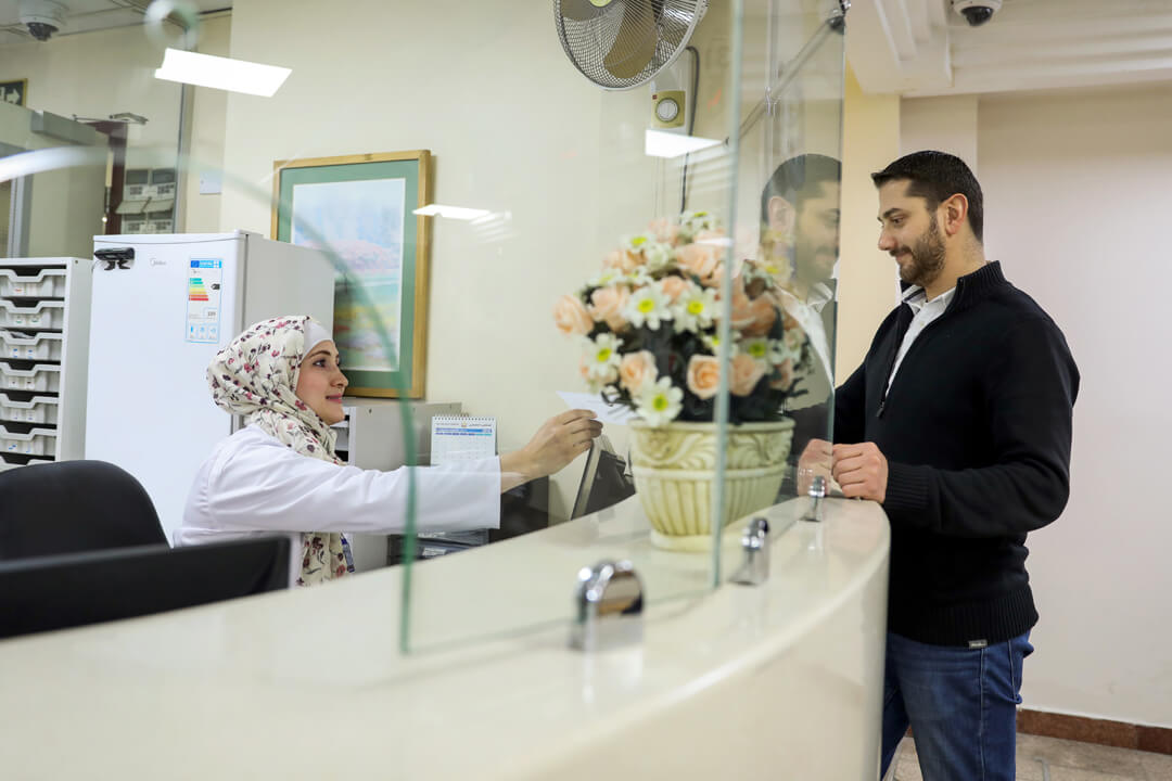
Nuclear medicine services are available to outpatients as well as inpatients during the following hours:
Saturday to Wednesday 8:30 am to 7:00 pm Thursday 8:30am to 2:00 pm


Her Majesty Rania Al-Abdullah
Jordan
I would like to highly express my admiration for your distinguished efforts that contributes to the development of the medical sector and to our county. I am pleased to express to all the staff at this outstanding medical institute, my great satisfaction and joy of the superior level the hospital achieved. I applaud for these great efforts and I support you to proceed for further achievements

Dr. Nabil Al–Sharif
Former Minister of Media & Communication in Jordan
Without exaggeration, I say that this medical institute provides great services around the clock to the citizens of Jordan, Arab patients and Jordan's visitors. The Specialty Hospital name in people's minds is connected to care and trust, which is a pride for our country and our medical institutes.

Fatima Khaled Al Bashir - Sudan
The Wife and first cousin of Sudanese president
Thank you very much Specialty Hospital for the warm welcoming and the good treatment we found, and we noticed the good reputation of the hospital's staff from administrative to doctors and we ask God to bless them

Dr. Gabriella Vicuna - USA
Medical Tourism Association
It was our greatest pleasure and honor to visit you during the MTA FAM tour. We are extremely impressed with the great quality, technology and talented physicians we found at Specialty Hospital. From the moment you walk through the doors of Specialty Hospital, one feels warmth and hospitality of this great facility

Dr. Ahmed Al-Asbahi – Yemen
Member of Shura Council of the Republic of Yemen
I didn't find the words to express the state of progress I have seen at the Specialty Hospital, but I would like to say that the secret of this progress is referred to the man expressed by HM King Hussein, may God bless him and grant him peace, he said: "Human is the most precious thing we have”. This statement has been scientifically translated into the existence of qualified competencies in various fields especially what I have seen at the Specialty Hospital, I witnessed the stages of its development. And here I am today witnessing the reality of dealing directly with the excellence in its management, wonderful nursing and advanced medicine.

Dr. Tabita Boutros - Sudan
Former Minister of Water and Dams - Sudan
I was very pleased to visit your hospital, and I came to receive medical care and general checkups. This is an opportunity to say thank you so much for everything you have done for me
Dr. Cris Mandy - USA
Medical Provider
Your hospital is excellent and state of the art. It is equivalent to our best hospitals in America. Your commitment to medical education is evident and serves as an investment into the future of health care in Jordan.We appreciate the great care you have provide our ill and injured American patients.

Tukunobu kita - Japan
Kyushu University
We visited this hospital as field research on medical tourism. The doctors kindly introduced the facilities and explained to us everything. We learned that Specialty Hospital has well-trained staff and good equipment as well as sophisticated systems. It has strong appeal for overseas patients

Anamaria Trotingher - Romania
Patient
I like to thanks Dr. Basheer Abu Halalh and all the crew at Specialty Hospital , I was in the royal suites, the hospital is perfect and super cleanthe crew is helpful and well educated. I want to thank every one who helped me and made my stay there as perfect as possible
LT. Chire Reilly - USA
White House Medical Unit
Thank you for your welcoming hospitality and for showing us your beautiful hospital, we look forward to working with you now and in the future

Dr. Anna - Netherlands
Medical Provider
Thank you so much for all the support, help and cooperation in the last few months. Thank you for all the care for our patients and for helping me doing my job over here in Jordan
Mike Williams - Louisiana
Patient
I would like to say a big thank you to all the staff at Specialty Hospital. The care I received here was great and I highly recommend it to everyone.
Dr. Mandy - USA
Medical Provider
Your emergency department was fantastic. In 30 years of practicing medicine in the United States I have never been able to get X-rays, an ultrasound and an MRI done on one of my patients in less than 4 hours. Your hospital's efficiency and timeliness and exemplary and set the standard.Our patient went to the operating theater and had his injury successfully repaired. He is doing well thanks to the great care he received at Specialty Hospital.
Artom Neekono - USA
Medical Provider
To the men and women that make up the wonderful staff of Specialty Hospital, thank you for the care and professionalism that you have consistently provided to all that came through you doors. You have provided a piece of mind knowing that our patients will have the best care possible in what would be normally be a very difficult situation. Thank you from the bottom of my heart

HRH Princess Muna Al-Hussain
Jordan
I am pleased to express my deep appreciation and admiration for the level of achievements you have reached in the medical field for our dear country. wish you success and good luck under the Patronage of His Majesty King Abdullah II Ibn Al Hussein